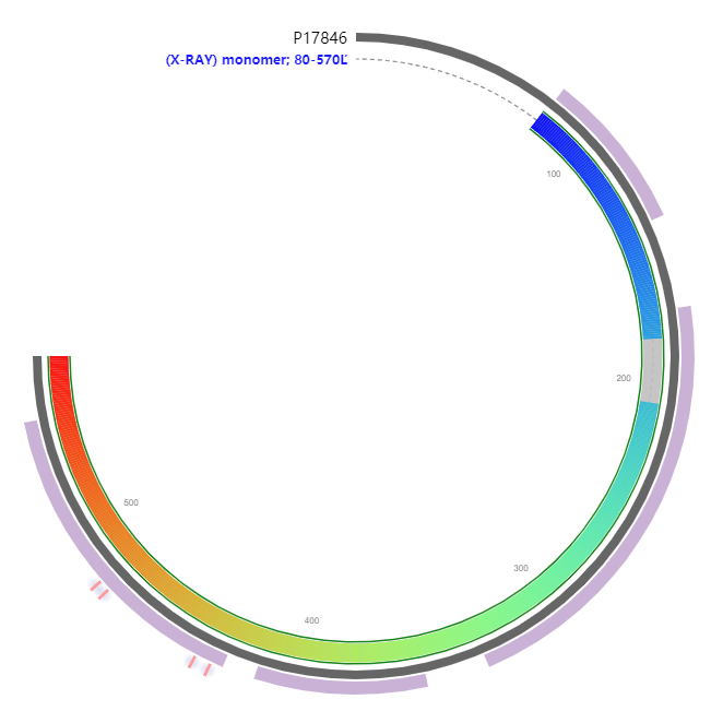
Component of the sulfite reductase complex that catalyzes the 6-electron reduction of sulfite to sulfide. This is one of several activities required for the biosynthesis of L-cysteine from sulfate.
MSEKHPGPLVVEGKLTDAERMKHESNYLRGTIAEDLNDGLTGGFKGDNFLLIRFHGMYQQ
DDRDIRAERAEQKLEPRHAMLLRCRLPGGVITTKQWQAIDKFAGENTIYGSIRLTNRQTF
QFHGILKKNVKPVHQMLHSVGLDALATANDMNRNVLCTSNPYESQLHAEAYEWAKKISEH
LLPRTRAYAEIWLDQEKVATTDEEPILGQTYLPRKFKTTVVIPPQNDIDLHANDMNFVAI
AENGKLVGFNLLVGGGLSIEHGNKKTYARTASEFGYLPLEHTLAVAEAVVTTQRDWGNRT
DRKNAKTKYTLERVGVETFKAEVERRAGIKFEPIRPYEFTGRGDRIGWVKGIDDNWHLTL
FIENGRILDYPARPLKTGLLEIAKIHKGDFRITANQNLIIAGVPESEKAKIEKIAKESGL
MNAVTPQRENSMACVSFPTCPLAMAEAERFLPSFIDNIDNLMAKHGVSDEHIVMRVTGCP
NGCGRAMLAEVGLVGKAPGRYNLHLGGNRIGTRIPRMYKENITEPEILASLDELIGRWAK
EREAGEGFGDFTVRAGIIRPVLDPARDLWD579

| PMID | Title & Author | Abstract | Year | |
| 0 | 2670946 | Characterization of the cysJIH regions of Salmonella typhimurium and Escherichia coli B. DNA sequences of cysI and cysH and a model for the siroheme-Fe4S4 active center of sulfite reductase hemoprotein based on amino acid homology with spinach nitrite reductase.Ostrowski J, Wu JY, Rueger DC, Miller BE, Siegel LM, Kredich NM | The hemoprotein component of Salmonella typhimurium sulfite reductase (NADPH) (EC 1.8.1.2) was purified to homogeneity from cysJ266, a mutant strain lacking sulfite reductase flavoprotein. The siroheme- and Fe4S4-containing enzyme was isolated as a monomeric 63-kDa polypeptide and consisted of a mixture of unligated enzyme and a complex with sulfite. Following reduction with 5'-deazaflavin-EDTA and reoxidation, the complex was converted to the uncomplexed, high spin ferri-siroheme state seen previously with Escherichia coli sulfite reductase hemoprotein preparations. The S. typhimurium hemoprotein exhibited catalytic and physical properties identical to the hemoprotein prepared by urea dissociation of E. coli sulfite reductase holoenzyme and was fully competent in reconstituting NADPH-sulfite reductase activity when combined with excess purified sulfite reductase flavoprotein. The DNA sequences of cysI and cysH from S. typhimurium and E. coli B were determined and, together with previously reported data, confirmed the organization of this region as promoter-cysJ-cysI-cysH with all three genes oriented in the same direction from the promoter. Molecular weights deduced for the cysI-encoded sulfite reductase hemoprotein and for the cysH-encoded 3'-phosphoadenosine 5'-phosphosulfate sulfotransferase were approximately 64,000 and 28,000, respectively. Comparison of the deduced amino acid sequence of sulfite reductase hemoprotein with that of spinach nitrite reductase (Back, E., Burkhart, W., Moyer, M., Privalle, L., and Rothstein, S. (1988) Mol. Gen. Genet. 212, 20-26), which also contains siroheme and an Fe4S4 cluster, showed two groups of cysteine-containing sequences with the structures Cys-(X)3-Cys and Cys-(X)5-Cys, which are homologous in the two enzymes and are postulated to provide the ligands of the Fe4S4 cluster in both proteins. From these sequences and from crystallographic (McRee, D. E., Richardson, D. C., Richardson, J. S., and Siegel, L. M. (1986) J. Biol. Chem. 261, 10277-10281) and spectroscopic data in the literature, a model is proposed for the structure of the active center of these two enzymes. | 1989 |
| 1 | 10984484 | A simplifed functional version of the Escherichia coli sulfite reductase. | Escherichia coli sulfite reductase (SiR) is a large and soluble enzyme with an alpha(8)beta(4) quaternary structure. Protein alpha (or sulfite reductase flavoprotein) contains both FAD and FMN, whereas protein beta (or sulfite reductase hemoprotein (SiR-HP)) contains an iron-sulfur cluster coupled to a siroheme. The enzyme is set up to arrange the redox cofactors in a FAD-FMN-Fe(4)S(4)-Heme sequence to make an electron pathway between NADPH and sulfite. Whereas alpha spontaneously polymerizes, we have been able to produce SiR-FP60, a monomeric but fully active truncated version of it, lacking the N-terminal part (Zeghouf, M., Fontecave, M., Macherel, D., and Covès, J. (1998) Biochemistry 37, 6114-6123). Here we report the cloning, overproduction, and characterization of the beta subunit. Pure recombinant SiR-HP behaves as a monomer in solution and is identical to the native protein in all its characteristics. Moreover, we demonstrate that the combination of SiR-FP60 and SiR-HP produces a functional 1:1 complex with tight interactions retaining about 20% of the activity of the native SiR. In addition, fully active SiR can be reconstituted by incubation of the octameric sulfite reductase flavoprotein with recombinant SiR-HP. Titration experiments and spectroscopic properties strongly suggest that the holoenzyme should be described as an alpha(8)beta(8) with equal amounts of alpha and beta subunits and that the alpha(8)beta(4) structure is probably not correct. | 2000 |
| 2 | 2158792 | Molecular cloning of the cys genes (cysC, cysD, cysH, cysI, cysJ, and cysG) responsible for cysteine biosynthesis in Escherichia coli K-12.H Tei, K Murata, A Kimura | The cysCDHIJ gene region at 59 min and the cysG gene at 74 min on the chromosome of Escherichia coli K-12 were cloned. The ability of these cloned genes to complement mutants was analyzed and their restriction maps were determined. The following results were obtained. First, the cysCDHIJ regulon could be separated into cysCD and cysHIJ regions. Second, the cysG gene was quite different from the cysI and cysJ genes. | 1990 |
Zeghouf, M. A Simplifed Functional Version of the Escherichia coli Sulfite Reductase[J]. Journal of Biological Chemistry, 2000, 275(48):37651-37656.