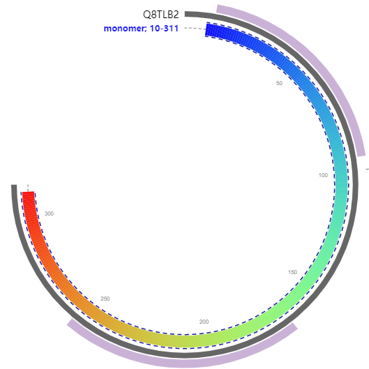
Heterodisulfide reductase (Hdr), is an iron-sulfur protein which in anaerobic methanogenic archaea catalyzes the reduction of the disulphide bond between coenzyme M and coenzyme B and is coupled to methane formation
MKAIESIPDRKLLLFKSCMVGQEYPGIETATSYVFDRLGVDYCINDEQSCCTGIGHYTDV
FEGLTTAAIAARNFAVARKCGYPNITCLCSTCYAINKDACELLNTNDGVREKVNSIFREK
GFDDLVYEKDSMNPRTNIYHAVEVLLSKVEKIREEIKFDFPGVKAASHHACHYYKVKYLD
VIGNPENPQLIDTIAEACGASPVRWYEDRTLTCGMGFSQLHLNKSTSLQVTKTKLDSLQR
AGVELMIHMCPNCHIQYDRYQPVIEKEFGVEYDMVHMNIAQFVALSMGADPYKVCGFQTH
SVPLEGFLEKTGII319

| PMID | Title & Author | Abstract | Year | |
| 0 | 7925445 | The heterodisulfide reductase from Methanobacterium thermoautotrophicum contains sequence motifs characteristic of pyridine-nucleotide-dependent thioredoxin reductases.R Hedderich 1, J Koch, D Linder, R K Thauer | The genes hdrA, hdrB and hdrC, encoding the three subunits of the iron-sulfur flavoprotein heterodisulfide reductase, have been cloned and sequenced. HdrA (72.19 kDa) was found to contain a region of amino acid sequence highly similar to the FAD-binding domain of pyridine-nucleotide-dependent disulfide oxidoreductases. Additionally, 110 amino acids C-terminal to the FAD-binding consensus, a short polypeptide stretch (VX2CATID) was detected which shows similarity to the region of thioredoxine reductase that contains the active-site cysteine residues (VX2CATCD). These findings suggest that HdrA harbors the site of heterodisulfide reduction and that the catalytic mechanism of the enzyme is similar to that of pyridine-nucleotide-dependent thioredoxin reductase. HdrA was additionally found to contain four copies of the sequence motif CX2CX2CX3C(P), indicating the presence of four [4Fe-4S] clusters. Two such sequence motifs were also present in HdrC (21.76 kDa), the N-terminal amino acid sequence of which showed sequence similarity to the gamma-subunit of the anaerobic glycerol-3-phosphate dehydrogenase of Escherichia coli. HdrC is therefore considered to be an electron carrier protein that contains two [4Fe-4S] clusters. HdrB (33.46 kDa) did not show sequence similarity to other known proteins, but appears to possess a C-terminal hydrophobic alpha-helix that might function as a membrane anchor. Although hdrB and hdrC are juxtaposed, these genes are not near hdrA. | 1994 |
| 1 | 19703284 | Extending the models for iron and sulfur oxidation in the extreme acidophile Acidithiobacillus ferrooxidans.Raquel Quatrini 1, Corinne Appia-Ayme, Yann Denis, Eugenia Jedlicki, David S Holmes, Violaine Bonnefoy | Background: Acidithiobacillus ferrooxidans gains energy from the oxidation of ferrous iron and various reduced inorganic sulfur compounds at very acidic pH. Although an initial model for the electron pathways involved in iron oxidation has been developed, much less is known about the sulfur oxidation in this microorganism. In addition, what has been reported for both iron and sulfur oxidation has been derived from different A. ferrooxidans strains, some of which have not been phylogenetically characterized and some have been shown to be mixed cultures. It is necessary to provide models of iron and sulfur oxidation pathways within one strain of A. ferrooxidans in order to comprehend the full metabolic potential of the pangenome of the genus.Results: Bioinformatic-based metabolic reconstruction supported by microarray transcript profiling and quantitative RT-PCR analysis predicts the involvement of a number of novel genes involved in iron and sulfur oxidation in A. ferrooxidans ATCC23270. These include for iron oxidation: cup (copper oxidase-like), ctaABT (heme biogenesis and insertion), nuoI and nuoK (NADH complex subunits), sdrA1 (a NADH complex accessory protein) and atpB and atpE (ATP synthetase F0 subunits). The following new genes are predicted to be involved in reduced inorganic sulfur compounds oxidation: a gene cluster (rhd, tusA, dsrE, hdrC, hdrB, hdrA, orf2, hdrC, hdrB) encoding three sulfurtransferases and a heterodisulfide reductase complex, sat potentially encoding an ATP sulfurylase and sdrA2 (an accessory NADH complex subunit). Two different regulatory components are predicted to be involved in the regulation of alternate electron transfer pathways: 1) a gene cluster (ctaRUS) that contains a predicted iron responsive regulator of the Rrf2 family that is hypothesized to regulate cytochrome aa(3) oxidase biogenesis and 2) a two component sensor-regulator of the RegB-RegA family that may respond to the redox state of the quinone pool.Conclusion: Bioinformatic analysis coupled with gene transcript profiling extends our understanding of the iron and reduced inorganic sulfur compounds oxidation pathways in A. ferrooxidans and suggests mechanisms for their regulation. The models provide unified and coherent descriptions of these processes within the type strain, eliminating previous ambiguity caused by models built from analyses of multiple and divergent strains of this microorganism. | 2009 |
| 2 | 17929940 | A cysteine-rich CCG domain contains a novel [4Fe-4S] cluster binding motif as deduced from studies with subunit B of heterodisulfide reductase from Methanothermobacter marburgensis.Nils Hamann 1, Gerd J Mander, Jacob E Shokes, Robert A Scott, Marina Bennati, Reiner Hedderich | Heterodisulfide reductase (HDR) of methanogenic archaea with its active-site [4Fe-4S] cluster catalyzes the reversible reduction of the heterodisulfide (CoM-S-S-CoB) of the methanogenic coenzyme M (CoM-SH) and coenzyme B (CoB-SH). CoM-HDR, a mechanistic-based paramagnetic intermediate generated upon half-reaction of the oxidized enzyme with CoM-SH, is a novel type of [4Fe-4S]3+ cluster with CoM-SH as a ligand. Subunit HdrB of the Methanothermobacter marburgensis HdrABC holoenzyme contains two cysteine-rich sequence motifs (CX31-39CCX35-36CXXC), designated as CCG domain in the Pfam database and conserved in many proteins. Here we present experimental evidence that the C-terminal CCG domain of HdrB binds this unusual [4Fe-4S] cluster. HdrB was produced in Escherichia coli, and an iron-sulfur cluster was subsequently inserted by in vitro reconstitution. In the oxidized state the cluster without the substrate exhibited a rhombic EPR signal (gzyx = 2.015, 1.995, and 1.950) reminiscent of the CoM-HDR signal. 57Fe ENDOR spectroscopy revealed that this paramagnetic species is a [4Fe-4S] cluster with 57Fe hyperfine couplings very similar to that of CoM-HDR. CoM-33SH resulted in a broadening of the EPR signal, and upon addition of CoM-SH the midpoint potential of the cluster was shifted to values observed for CoM-HDR, both indicating binding of CoM-SH to the cluster. Site-directed mutagenesis of all 12 cysteine residues in HdrB identified four cysteines of the C-terminal CCG domain as cluster ligands. Combined with the previous detection of CoM-HDR-like EPR signals in other CCG domain-containing proteins our data indicate a general role of the C-terminal CCG domain in coordination of this novel [4Fe-4S] cluster. In addition, Zn K-edge X-ray absorption spectroscopy identified an isolated Zn site with an S3(O/N)1 geometry in HdrB and the HDR holoenzyme. The N-terminal CCG domain is suggested to provide ligands to the Zn site. | 2007 |
| 3 | 24037219 | Advanced electron paramagnetic resonance on the catalytic iron-sulfur cluster bound to the CCG domain of heterodisulfide reductase and succinate: quinone reductase.Alistair J Fielding 1, Kristian Parey, Ulrich Ermler, Silvan Scheller, Bernhard Jaun, Marina Bennati | Heterodisulfide reductase (Hdr) is a key enzyme in the energy metabolism of methanogenic archaea. The enzyme catalyzes the reversible reduction of the heterodisulfide (CoM-S-S-CoB) to the thiol coenzymes M (CoM-SH) and B (CoB-SH). Cleavage of CoM-S-S-CoB at an unusual FeS cluster reveals unique substrate chemistry. The cluster is fixed by cysteines of two cysteine-rich CCG domain sequence motifs (CX31-39CCX35-36CXXC) of subunit HdrB of the Methanothermobacter marburgensis HdrABC complex. We report on Q-band (34 GHz) (57)Fe electron-nuclear double resonance (ENDOR) spectroscopic measurements on the oxidized form of the cluster found in HdrABC and in two other CCG-domain-containing proteins, recombinant HdrB of Hdr from M. marburgensis and recombinant SdhE of succinate: quinone reductase from Sulfolobus solfataricus P2. The spectra at 34 GHz show clearly improved resolution arising from the absence of proton resonances and polarization effects. Systematic spectral simulations of 34 GHz data combined with previous 9 GHz data allowed the unambiguous assignment of four (57)Fe hyperfine couplings to the cluster in all three proteins. (13)C Mims ENDOR spectra of labelled CoM-SH were consistent with the attachment of the substrate to the cluster in HdrABC, whereas in the other two proteins no substrate is present. (57)Fe resonances in all three systems revealed unusually large (57)Fe ENDOR hyperfine splitting as compared to known systems. The results infer that the cluster's unique magnetic properties arise from the CCG binding motif. | 2013 |
Ehrenfeld N , Levicán, Gloria, Parada P . Heterodisulfide Reductase from Acidithiobacilli is a Key Component Involved in Metabolism of Reduced Inorganic Sulfur Compounds[J]. Advanced Materials Research, 2013, 825:194-197.