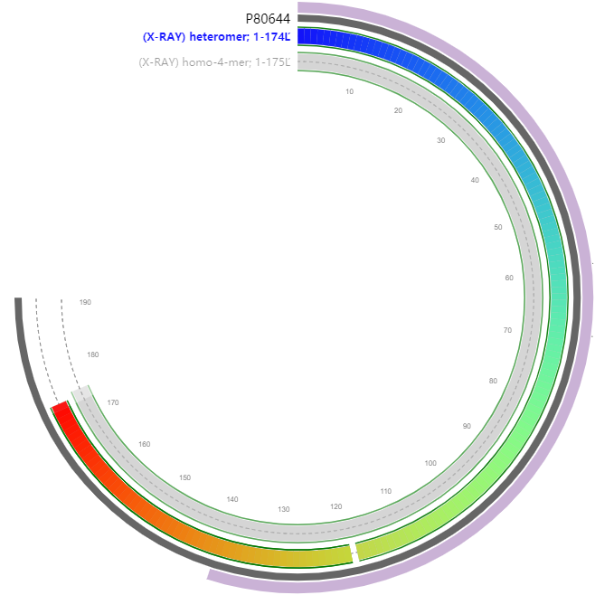
Probably forms a two-component reduced flavin mononucleotide-dependent monooxygenase by binding to SsuD. Required for growth on aliphatic sulfonates or methionine but not arylsulfonates
MRVITLAGSPRFPSRSSSLLEYAREKLNGLDVEVYHWNLQNFAPEDLLYARFDSPALKTF
TEQLQQADGLIVATPVYKAAYSGALKTLLDLLPERALQGKVVLPLATGGTVAHLLAVDYA
LKPVLSALKAQEILHGVFADDSQVIDYHHRPQFTPNLQTRLDTALETFWQALHRRDVQVP
DLLSLRGNAHA194

| PMID | Title & Author | Abstract | Year | |
| 0 | 10480865 | Characterization of a two-component alkanesulfonate monooxygenase from Escherichia coli.Eichhorn E, van der Ploeg JR, Leisinger T | The sequence of the dsr gene region of the phototrophic sulfur bacterium Chromatium vinosum D (DSMZ 180) was determined to clarify the in vivo role of 'reverse' sirohaem sulfite reductase. The dsrAB genes encoding dissimilatory sulfite reductase are part of a gene cluster, dsrABEFHCMK, that encodes four small, soluble proteins (DsrE, DsrF, DsrH and DsrC), a transmembrane protein (DsrM) with similarity to haem-b-binding polypeptides and a soluble protein (DsrK) resembling [4Fe-4S]-cluster-containing heterodisulfide reductase from methanogenic archaea. Northern hybridizations showed that expression of the dsr genes is increased by the presence of reduced sulfur compounds. The dsr genes are not only transcribed from a putative promoter upstream of dsrA but primary transcripts originating from (a) transcription start site(s) downstream of dsrB are also formed. Polar insertion mutations immediately upstream of dsrA, and in dsrB, dsrH and dsrM, led to an inability of the cells to oxidize intracellularly stored sulfur. The capability of the mutants to oxidize sulfide, thiosulfate and sulfite under photolithoautotrophic conditions was unaltered. Photoorganoheterotrophic growth was also unaffected. 'Reverse' sulfite reductase and DsrEFHCMK are, therefore, not essential for oxidation of sulfide or thiosulfate, but are obligatory for sulfur oxidation. These results, together with the finding that the sulfur globules of C. vinosum are located in the extracytoplasmic space whilst the dsr gene products appear to be either cytoplasmic or membrane-bound led to the proposal of new models for the pathway of sulfur oxidation in this phototrophic sulfur bacterium. | 1999 |
| 1 | 29979040 | Functional Evaluation of the π-Helix in the NAD(P)H:FMN Reductase of the Alkanesulfonate Monooxygenase System.Jonathan M Musila , Dianna L Forbes , Holly R Ellis | A subgroup of enzymes in the NAD(P)H:FMN reductase family is comprised of flavin reductases from two-component monooxygenase systems. The diverging structural feature in these FMN reductases is a π-helix centrally located at the tetramer interface that is generated by the insertion of an amino acid in a conserved α4 helix. The Tyr insertional residue of SsuE makes specific contacts across the dimer interface that may assist in the altered mechanistic properties of this enzyme. The Y118F SsuE variant maintained the π-π stacking interactions at the tetramer interface and had kinetic parameters similar to those of wild-type SsuE. Substitution of the π-helical residue (Tyr118) to Ala or Ser transformed the enzymes into flavin-bound SsuE variants that could no longer support flavin reductase and desulfonation activities. These variants | |
| existed as dimers and could form protein-protein interactions with SsuD even though flavin transfer was not sustained. The ΔY118 SsuE variant was flavin-free as purified and did not undergo the tetramer to dimer oligomeric shift with the addition of flavin. The absence of desulfonation activity can be attributed to the inability of ΔY118 SsuE to promote flavin transfer and undergo the requisite oligomeric changes to support desulfonation. Results from these studies provide insights into the role of the SsuE π-helix in promoting flavin transfer and oligomeric changes that support protein-protein interactions with SsuD. | 2018 | |||
| 2 | 27806563 | Transformation of a Flavin-Free FMN Reductase to a Canonical Flavoprotein through Modification of the π-Helix.Jonathan M Musila , Holly R Ellis | The flavin reductase of the alkanesulfonate monooxygenase system (SsuE) contains a conserved π-helix located at the tetramer interface that originates from the insertion of Tyr118 into helix α4 of SsuE. Although the presence of π-helices provides an evolutionary gain of function, the defined role of these discrete secondary structures remains largely unexplored. The Tyr118 residue that generated the π-helix in SsuE was substituted with Ala to evaluate the functional role of this distinctive structural feature. Interestingly, generation of the Y118A SsuE variant converted the typically flavin-free enzyme to a flavin-bound form. Mass spectrometric analysis of the extracted flavin gave a mass of 457.11 similar to that of the FMN cofactor, suggesting the Y118A SsuE variant retained flavin specificity. The Y118A SsuE FMN cofactor was reduced with approximately 1 equiv of NADPH in anaerobic titration experiments, and the flavin remained bound following reduction. Although reactivity of the reduced flavin with oxygen was slow in NADPH oxidase assays, the variant supported electron transfer to ferricyanide. In addition, there was no measurable sulfite product in coupled assays with the Y118A SsuE variant and SsuD, further demonstrating that flavin transfer was no longer supported. The results from these studies suggest that the π-helix enables SsuE to effectively utilize flavin as a substrate in the two-component monooxygenase system and provides a foundation for further studies aimed at evaluating the functional properties of the π-helix in SsuE and related two-component flavin reductase enzymes. | 2016 |
Eichhorn E , Van d P J R , Leisinger T . Characterization of a Two-component Alkanesulfonate Monooxygenase from Escherichia coli[J]. Journal of Biological Chemistry, 1999, 274(38):26639-26646.